by Amanda J. Doolin-Carver, OTDR/L, ATP
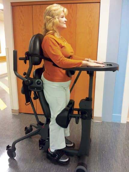
 In static standing, standing frames may be appropriate based on the patient’s ability to maintain head control.Approximately 12,000 new spinal cord injuries are reported each year in the United States. As many as 306,000 Americans may be living with a spinal cord injury. These injuries include any insult that results in change in normal motor, sensory, or autonomic function and can be used to describe tetraplegia (inclusion of four extremities) or paraplegia (injury in thoracic, lumbar, or sacral spinal cord).1 Spinal cord injuries also can be defined as either complete, no motor or sensory function in the sacral segments S4-S5, or incomplete, either sensory function is preserved at the S4-S5 segment or motor function is preserved below the neurological level of injury.2 Any spinal cord injury dysfunction can result in the person’s inability to stand or bear weight through the lower extremities independently.
SECONDARY COMPLICATIONS POST-SCI
After a person incurs a spinal cord injury, that individual’s life is changed in a number of ways. Of course, the most obvious changes are motor paralysis and loss of sensory function; but they also have to face all of the secondary complications. These complications include but are not limited to osteoporosis, spasticity, risk of pressure sores, urinary dysfunction, and insufficient cardiac output. Each secondary complication leads to complications of its own. For instance, osteoporosis leads to fractures that decrease a person’s independence and function. Osteoporosis is a condition in which the bone loses density and becomes more fragile and likely to fracture. It occurs when the body fails to form enough new bone, when too much existing bone is reabsorbed by the body, or both.3 Individuals with spinal cord injury have an altered skeletal structure because of the altered pattern of loading of the lower extremities, which leads to loss of 50% to 60% of their bone mineral density.4,5 It is documented that loss is greatest in the first year after spinal cord injury, but continues throughout life with no intervention. A monthly loss of 2% cortical bone and 4% trabecular bone for the first year after spinal cord injury has been documented.4 Maintaining or returning the normal pattern of loading the lower extremities through weight bearing can attenuate the bone loss in individuals with spinal cord injury, thereby significantly decreasing the risk of osteoporosis.
Other complications for persons with spinal cord injury include sensory loss, which leads to the complication of pressure ulcers. Pressure ulcers require extensive, expensive healing, sometimes including surgical repair, which leads to lengthy bedrest and increased use of caregivers. Pressure ulcers are a significant risk for persons with a spinal cord injury. They are caused by a person’s inability to move weight so that the blood can nourish the tissues. As many as 33% of spinal cord injured persons will incur a pressure ulcer in their lifetime.
A third significant complication is spasticity. Spasticity decreases a person’s ability to independently transfer as well as independently complete ADLs. Spasticity is caused by a lack of inhibition, allowing excessive contraction of the muscle, and can be elicited or inhibited by certain stretch reflexes or positions. All secondary complications lead to loss of function, loss of independence, need for medical intervention, and increased mortality.
ESTABLISHING A STANDING PROGRAM
It is the duty of rehabilitation personnel to decrease the risk of secondary complications following spinal cord injury. One way in which the secondary complications can be managed is through a standing program. The benefits of standing after spinal cord injury are well known. Standing has been shown to decrease urinary calculi, decrease bone demineralization, decrease spasticity, increase pressure relief, and increase morale and quality of life, to name only a few of the benefits. Increased urinary calculi pose a risk of infection, increase risk of kidney stones, and demonstrate calcium loss from the bones. Studies over the years of persons with spinal cord injury and noninjured persons have demonstrated that bedrest increases urinary calculi and that passive standing decreases urinary calculi.6,7
Extensive studies have been completed demonstrating the bone demineralization with nonuse of the lower extremities. Regular standing provides sufficient mechanical loads to maintain bone mineral density. In as little as 30 minutes per day, bone mineral density loss can be decreased significantly or even stopped completely.4 Spasticity can be reduced through standing by providing prolonged stretch to the skeletal muscles.8 Pressure relief can be obtained by the change of position provided by standing. The most beneficial method of prevention is complete offloading of the pressure points, which can be obtained by standing. Quality of life is increased by the decreased risk of secondary complications during standing, the achievement of standing, and increased level of function while standing. Although multiple other benefits exist, these benefits of standing are the most well known and well documented.
ASSESSING THE OPTIONS
Many options are available for standing, including standing frames, tilt tables, dynamic standing frames, and neuromuscular electrical stimulation (NMES). To determine an individual’s best choice it is necessary to know the options. Standing frames and tilt tables are forms of static standing that provide support and allow a person to remain in a standing position for a prolonged period. To determine which is the best form of support to maintain a static position, the person’s level of support needs should be addressed. If the person has the ability to maintain head control, a standing frame can be sufficient, but if a person requires increased head support or experiences orthostatic hypotension that would require slow upward movement and quick downward movement, then a tilt table should be used.
The location where a standing device will be used is another consideration in prescription. If it will be used in the home, a standing frame is more space efficient and easier to store than a tilt table. Transfer onto and off a standing frame is easier than the dependent slide that would be necessary for a tilt table. Some standing frames can be used from the wheelchair and no transfer is necessary. If the person has enough trunk strength and requires support only at the hips (which can be provided by a strap) and the knees (which is provided through block pads), a standing frame using the current wheelchair is the best option. Another consideration is the desired benefit of the standing program. Passive standing using a tilt table can have immediate effects on spasticity, although the effects of a single session are short-lived.8 Passive standing using a tilt table is shown to be more effective at decreasing extensor spasms than flexor spasms. Passive standing using standing frames has been shown to improve standing symmetry, reduce bone density loss, and decrease spasticity.6-8
WHAT DYNAMIC STANDING CAN OFFER
Other alternatives for standing programs include dynamic standing. The options in dynamic standing comprise dynamic standing frames, body weight supported systems, and NMES. When determining the best option between passive and dynamic standing programs, it is necessary to consider the ease of use, tolerance, and assistance level available for the person. Body weight supported systems and NMES require the expertise of a therapist for setup and guidance when using the systems. Body weight supported systems are also very expensive and usually require a significant amount of space or home modification for the mechanism. The dynamic standing programs not only incorporate weight bearing and muscle stretching, but also include electromyographic activity, which adds the advantage of cardiovascular, metabolic, and cellular changes to the musculature.8-10 Body weight supported training has been shown to decrease flexor spasms more effectively than passive standing.8 Considerations for the use of an NMES program include the requirement that the program be standing. Studies have shown no increased benefit over static standing when using NMES assisted cycling,8,10 and seated muscle contraction through NMES can generate large shear forces capable of fracture.5 Dynamic standing NMES programs provide the optimal compression and minimize shear forces to achieve the most effective use of time, highest amount of bone mineral density preservation, and highest rate of muscle modulation for decreasing spasticity.5,8-10 All of the above factors should be considered when choosing the most appropriate standing program.
TIMING RECOVERY
The final consideration when completing standing programs is the time requirement. Since bone mineral density decreases most significantly in the first year after injury, it is important to begin a standing program within the first 6 months after injury and continue the program for life. A static standing program should be completed 60 minutes to 3 hours per day, 5 days per week, to decrease bone mineral density loss and decrease muscle spasticity. A dynamic standing program using a body weight supported system should be completed 3 days to 5 days per week for 60 minutes. A dynamic standing program using NMES can be effective in as little as three sessions per week, lasting 45 minutes each.
Static and dynamic standing programs can each be beneficial for an individual’s quality of life, and achieve many medical benefits. Since no program is beneficial without compliance, choosing the correct fit for each person is very important to achieve the optimal outcome.
References
1. http://emedicine.medscape.com/article/322480-overview
2. American Spinal Injury Association, Standard Neurological Classification of Spinal Cord Injury
3. http://www.ncbi.nlm.nih.gov/pubmedhealth/PMH0001400/
4. Dionyssiotis Y, Lyritis GP, Mavrogenis AF, Papagelopoulos PJ. Factors influencing bone loss in paraplegia. Hippokratia. 2011;15:54-59.
5. McHenry C, Shields R. A biomechanical analysis of exercise in standing, supine, and seated positions: implications for individuals with spinal cord injury. J Spinal Cord Med. 2012;35(3):140-147.
6. Leo K. The effects of passive standing. Paraplegia News. November 1985.
7. Newman M, Barker K. The effect of supported standing in adults with upper motor neurone disorders: a systematic review. Clin Rehabil. May 29, 2012. Epub ahead of print.
8. Adams M, Hicks A. Comparison of the effects of body-weight-supported treadmill training and tilt-table standing on spasticity in individuals with chronic spinal cord injury. J Spinal Cord Med. 2011; 34(5):488-494.
9. Kawashima N, Yano H, Ohta Y, Nakazawa K. Stretch reflex modulation during imposed static and dynamic hip movements in standing humans. Exp Brain Res. 2006;174:342-350.
10. Dudley-Javoroski S, Saha PK, Liang G, Li C, Gao Z, Shields RK. High dose compressive loads attenuate bone mineral loss in humans with spinal cord injury. Osteoporos Int. 2012;23:2335-2346.
Amanda J. Doolin-Carver, OTDR/L, ATP, received her bachelor of occupational therapy from Mount Mary College, Milwaukee, in 2001, then earned a doctorate of occupational therapy from Rocky Mountain University of Health Professions in 2008. She has been employed for 10 years at Memorial Medical Center Regional Rehabilitation Center, Springfield, Ill, and is currently at a master occupational therapist level working on the spinal cord injury team and the outpatient wheelchair clinic. For more information, contact [email protected].