Rehabilitative ultrasound imaging (RUSI) is a new modality that a physical therapist can use to retrain muscles. It is often utilized to treat patients with low back pain, and is defined as a procedure used by a physical therapist to evaluate muscles and related soft tissue morphology as it relates to behavior during physical therapy tasks using ultrasound imaging.1
For more than 10 years, RUSI has been used in physical therapy research. Those researchers who use RUSI report that they have discovered the multifidus muscles do not spontaneously recover after a back injury.2 Further studies showed a significant interaction between chronic low back pain and the ability to contract the multifidus muscles at a specific lumbar spinal segment.3 This study also concluded that a patient had to be able to isolate the transversus abdominis muscle (TrA) in order to activate the multifidus muscles.3 Often patients use their internal and external oblique muscles to control their abdominal region and the long lumbar extensors to move their lumbar spine. These studies point out that altered motor control plays a role in the pain cycle. The physical therapist and the patient can visualize those altered movement patterns, and the therapist can instruct the patient about how to change these patterns by using RUSI.
There is also now a clinical model of RUSI available, which is easy for a physical therapist to use. It only takes the use of a couple of buttons to be able to view the patient’s deep core muscles and lumbar multifidus muscles. Therapists need education, however, to understand what they are viewing. Enough ultrasound gel also must be used for sound wave transmission. Using RUSI, the therapist can view the layers of the abdominal wall including the external oblique muscle, internal oblique muscle, and a line for the transverse abdominis muscle. They can see the deep and superficial multifidus muscles as well. The muscle activation pattern can be visualized by using RUSI. This pattern demonstrates whether the patient is able to isolate the TrA. The correct muscle activation is a corseting action of the lowest layer of muscle. A large global picture using all the abdominal muscles to create the contraction is the incorrect activation. This is the typical activation pattern that patients with low back pain use.
Complementing Modalities to Manage pain
Sometimes the patient is in too much pain to relax this global pattern of muscle activation that may also activate the erector spinal muscles. When this happens, the therapist has several choices of modalities. Although decisions in treatment can vary from therapist to therapist when treating this patient population, among the modality options available to help treat low back pain are heat and cold therapy. While used in the RICE protocol to treat acute injuries, heat and cold therapy can be introduced to patients with low back pain and ultimately serve as a simple strategy for independent symptom management at home. A decrease in muscle spasms and increase in relaxation prior to stretching can be a benefit yielded by the use of moist hot packs. Heat also can provide a direct line to pain management in more centralized pain states, dilating blood vessels and improving oxygen flow and circulation to promote healing. As an adjunctive modality, cryotherapy can be used to control pain, constricting blood vessels to reduce swelling, or numbing injured tissue by slowing the nerve impulse. Contraindications in heat therapy include impaired sensation, thermal injury, circulatory issues, DVT, infections, malignant tumors, as well as sensitivity to heat. Cold therapy contraindications dictate that the therapy should not be used in patients with rheumatoid arthritis, Raynaud’s syndrome, cold allergic reaction, certain cardiac conditions, and impaired sensation.
An additional choice of modality for deep tissue treatment is the low-level cold laser, which promotes tissue healing at a cellular level and aligns the collagen fibers to improve scar tissue mobility. Contraindications for the low-level cold laser are cancer, area of hemorrhage, over pacemaker, and pregnancy. Certain facilities also have noted that blending conventional ultrasound with e-stim has presented positive outcomes when addressing flare-ups of chronic low back pain resulting from muscle spasms, and decreasing irritability of trigger points. In some clinics, iontophoresis may be used to decrease inflammation of a specific area on the low back.
Interferential electrical stimulation, TENS—a high frequency electrical stimulation built to be tolerated for longer periods of time and also allow for independent, home-based pain management—or topical creams that can be applied in the clinic or at home, are additional options that work to relax the overactive muscles. When these overactive muscles relax, then the activation pattern can be changed.
EDUCATING THE PATIENT
By using RUSI, the therapist begins to educate the patient about how to isolate the TrA. The patient can see the muscle contraction and adjust the amount of the force of the contraction by this visual feedback.
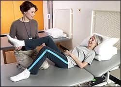
Deep relaxed breathing is the first step. The patient has to be able to breathe all the way down to the stomach. The best way to teach someone how to contract the TrA is in side-lying.4
1. Slowly draw the lower abdominal wall into the spine.
2. Slowly and gently contract the muscles that stop the flow of urine.
3. Draw the hip bones together, drawing the ASIS together.
4. Another way is to use tactile cueing with drawing the stomach away from the therapist’s hand.
The patient uses the feedback from the therapist and RUSI to modify their contraction. When comparing treatment of patients using the RUSI vs without the RUSI, therapists in general are advancing their patient too fast on back stabilization exercises. They quickly add arm raises to the patient’s home exercise program. When you view the TrA on RUSI, then you add single arm raise, frequently the patient cannot maintain the TrA contraction. Often patients take a couple of treatments using RUSI to be able to maintain the TrA with arm movement.
Another important fact in treating patients with RUSI is if the patient cannot isolate the TrA, then they will not be able to fire their multifidus muscles. The deep multifidus muscles stiffen the spine to allow movement. These muscles atrophy at the level of injury and do not recover unless they are specifically worked.2 This atrophy can be seen on MRI as fatty, white infiltrate in the multifidus muscles. Often the muscle tissue close to the spinous process is white, which is the deep multifidus muscle. This atrophy is most often seen at the L4-L5 segment, but can include multiple spinal segments.3 This muscle atrophy can be palpated by the therapist, on either side of the lumbar spinous processes. The palpating fingers sink in, like a hole. RUSI is an excellent tool to visualize these muscles. To activate these muscles, the patient is prone over a pillow and asked to breathe in/out and draw in the lower stomach. The therapist facilitates the contraction by tapping to obtain a muscle contraction under their fingers.
Then the therapist uses RUSI to see the muscles’ contraction. The sound head can be in a transverse direction to see both sides of the L5 multifidus muscle or can be alongside the spine to see the right multifidus muscles of L3-L4-L5. If the patient has less muscle bulk on palpation, fatty infiltrate on MRI, at L5, they will not be able to isolate the multifidus muscle at L5. The multifidus muscles at L2-L3 may contract more easily and with time the L5 will be activated. The patient is progressed to more functional positions.
Integrating Aquatics
Another way to change these altered motor patterns is through the use of the therapeutic pool. Through this approach, the patient is instructed, in the clinic, how to activate the TrA and then practices that activation with the therapist. The patient then uses the contraction they have been taught in the pool. The water assists the patient in determining whether they are indeed contracting the correct muscles. If their back begins to extend, they are not contracting the TrA. The water assists in proprioceptive feedback to promote spinal stability. The patient has less pain in the water and may be able to progress better than on land.
Physical therapy goals have been to change motor control patterns in patients to improve function and decrease pain. RUSI is a powerful tool for a physical therapist to use to educate patients about this change. When patients change their motor control patterns, they activate the small antigravity muscles such as the TrA and multifidus muscle to stabilize the spine, which decreases back pain and improves function. Studies have demonstrated the effectiveness of RUSI, and this may be an opportune time for an expansion of the technology and treatment. RM
Ellen O’Gorman, PT, has been a physical therapist for more than 30 years. She received her Bachelor of Science in Physical Therapy from St Louis University in 1983. She received a Masters in Health Science in 2005 from the University of Indianapolis. O’Gorman has been working in an outpatient setting for 15 years, specializing in lumbar and cervical spine patients. She was introduced to Rehabilitative Ultrasound Imaging in 2004. O’Gorman currently works for Premier Health at Miami Valley Hospital in Dayton, Ohio. For more information, contact [email protected].
References
1. Teyhen DS, Miltenberger CE, Deiters HM, et al. The use of ultrasound imaging of the abdominal drawing-in maneuver in subjects with low back pain. J Orthop Sports Phys Ther. 2005;35(6):346-355.
2. Hides JA, Stokes MJ, Saide M, et al. Evidence of lumbar multifidus muscle wasting ipsilateral to symptoms in patient with acute/subacute low back pain. Spine. 1994;19(2):165-177.
3. Hides J, Stanton W, Mendis MD, Sexton M. The relationship of transversus abdominis and lumbar multifidus clinical muscle tests in patients with chronic low back pain. Man Ther. 2011;16(6):573-7.
4. Lee DG. The Pelvic Girdle: An Approach to Examination and Treatment of the Lumbo-pelvic-hip Region. 3rd ed. Edinburgh: Churchill Livingstone; 2004.