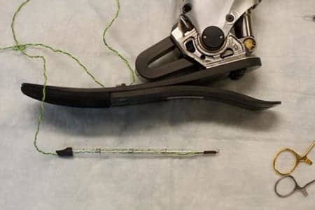
A new surgical approach developed at MIT aims to allow amputees to receive sensory feedback from their prosthetic limbs and improve their ability to control them. (Photo courtesy of Jose-Luis Olivares)
MIT researchers have devised a new surgical technique that coordinates a patient’s prosthetic limb with existing nerves and muscle grafts. They suggest this could ultimately enable the patient to sense and control the prosthetic.
In addition, the Massachusetts Institute of Technology (MIT) scientists note that this type of system could also help reduce the body’s rejection rate of prosthetic limbs, which is currently around 20%.
“We’re talking about a dramatic improvement in patient care,” says Hugh Herr, a professor of media arts and sciences and the senior author of the study, in a media release from MIT. “Right now there’s no robust neural method for a person with limb amputation to feel proprioceptive positions and forces applied to the prosthesis. Imagine how that would completely hinder one’s ability to move, to successfully balance, or to manipulate objects.”
The study appears in a recent issue of Science Robotics.
In their study, performed in rats, the team demonstrated that their technique generates muscle-tendon sensory feedback to the nervous system, which should be able to convey information about a prosthetic limb’s placement and the forces applied to it.
They did this by recreating the agonist-antagonist muscle relationships among the rats. In many amputees, the nerves that send signals to the amputated limb remain intact. The researchers decided to take advantage of those nerves by connecting them to muscle pairs grafted from another part of the body into the amputation site.
These grafts, which would be about 4 centimeters by 1.5 centimeters in humans, consist of a pair of muscles that work together just like natural muscles. When the brain sends signals instructing a limb to move, one of the grafted muscles will contract, and its agonist will extend. The agonist muscle then sends feedback to the brain about how much the muscle moved and the forces applied to it, the release explains.
After testing the muscle grafts in rats, the researchers found that when the rats contracted one muscle of the pair, the other muscle would move in the opposite way and send sensory information back to the brain.
“Using this framework, the patient will not have to think about how to control their artificial limb. When a patient imagines moving their phantom limb, signals will be sent through nerves to the surgically constructed muscle pairs. Implanted muscle electrodes will then sense these signals for the control of synthetic motors in the external prosthesis,” Herr states, in the release. “We think that because the brain is so good at remapping and it’s so plastic, it will quickly adapt to knowing how much it has to contract each muscle graft for natural prosthetic control.”
This type of feedback system should also allow people with a prosthetic arm, for example, to feel a torque applied to the prosthesis. “If you were to give a prosthetic-arm user a barbell to hold, they would actually feel the torque on the prosthetic wrist joint,” Herr adds.
This strategy could work for nearly any amputee, including people whose amputations were performed many years ago, the team continues.
“For almost any amputation scenario, as long as we have a little bit of the healthy nerve left, we can take that and put it into regenerative muscle grafts. We can harvest these muscle grafts from almost anywhere in the body, making this applicable to a large number of cases ranging from trauma to chronic pain,” concludes lead author Shriya Srinivasan, a graduate student in the Harvard-MIT Program in Health Sciences and Technology (HST), in the release.
[Source(s): Massachusetts Institute of Technology, Science Daily]


Great invention by MIT Scientists.