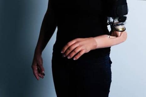
Shown here is an amputee fitted with an advanced arm prosthetic following TMSR surgery. (Photo credit: Irit Hacmun, Tel Aviv)
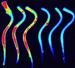
Using ultra-high field 7 Tesla fMRI, scientists illustrate how the brain re-maps motor and sensory pathways following a targeted motor and sensory reinnervation (TMSR) procedure in amputees.
TMSR is a surgical procedure performed on patients with amputations that reroutes residual limb nerves toward intact muscles and skin in order to fit them with a limb prosthesis allowing unprecedented control.
The scientists, from Ecole Polytechnique Fédérale de Lausanne, used fMRI to show how TMSR affects upper-limb representations in the brains of patients with amputations, in particular in primary motor cortex and the somatosensory cortex and regions processing more complex brain functions.
Results of their investigation were published in Brain, a media release explains.
The lab of Olaf Blanke at EPFL, in collaboration with Andrea Serino at the University Hospital of Lausanne and teams of clinicians and researchers in Switzerland and abroad have successfully mapped out these changes in the cortices of three patients with upper-limb amputations who had undergone TMSR and were proficient users of prosthetic limbs developed by Todd Kuiken and his group at the Rehabilitation Institute of Chicago.
Surprisingly, the study showed that motor cortex maps of the amputated limb were similar in terms of extent, strength, and topography to individuals without limb amputation, but they were different from patients with amputations that did not receive TMSR, but were using standard prostheses. This shows the unique impact of the surgical TMSR procedure on the brain’s motor map, the release explains.
The somatosensory maps showed that the brain had preserved its original topographical organization, although to a lesser degree than in healthy subjects. Moreover, when investigating the connections between upper-limb maps in both cortices, the researchers found normal connections in the TMSR patients, which were comparable with healthy controls. However, preservation of original mapping was again reduced in non-TMSR patients, showing that the TMSR procedure preserves strong functional connections between primary sensory and motor cortex.
The study also showed that TMSR is still in need of improvement: the connections between the primary sensory and motor cortex with the higher-level embodiment regions in fronto-parietal cortex were as weak in the TMSR patients as in the non-TMSR patients, and differed with respect to healthy subjects, the release continues.
This suggests that, despite enabling good motor performance, TMSR-empowered artificial limbs still do not move and feel like a real limb and are still not encoded by the patient’s brain as a real limb. The scientists conclude that future TMSR prosthetics should implement systematic somatosensory feedback linked to the robotic hand movements, enabling patients to feel the sensory consequences of the movements of their artificial limb.
The findings suggest that TMSR may counteract poorly adapted plasticity in the cortex after losing a limb. According to the authors, this may provide new insights into the nature and the reversibility of cortical plasticity in patients with amputations and its link to phantom limb syndrome and pain, the release concludes.
[Source(s): Ecole Polytechnique Fédérale de Lausanne, Science Daily]