A clear vision of patient goals and evidence-based treatment reinforces efficient and effective outcomes.
by Christina R. Lighthill, MOT, OTR, Erin E. Perez, PT, DPT, CBIS, and Keith B. McWilliams, MOT, OTR/L
 |
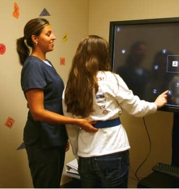
| Visual integrator addresses precision motor control, visual scanning, and attention. |
Many Americans who experience a brain injury (BI) suffer visual impairments as a result of the injury. Each year, an estimated 1.7 million Americans sustain a traumatic brain injury (TBI) and another 795,000 individuals suffer an acquired brain injury (ABI) from nontraumatic causes.1 Of those, as many as 40% experience some degree of neurological vision impairment.2 These deficits may often be overlooked or misunderstood; they may be missed initially when physicians or patients are focused on more pronounced problems and patients may lack awareness of their own vision loss.
For survivors of BI, visual dysfunction further complicates the recovery process. And, because visual input is closely tied to balance, these impairments can increase the risk of falling and sustaining a subsequent brain injury.
Examples of visual impairment symptoms include: making errors when copying written information, feeling unusually tired after completing a visual task, unusual posture or head tilt when reading or writing, difficulty negotiating around obstacles in a pathway, poor concentration, dizziness, headache, difficulty tracking moving objects, unusual eye rubbing, complaints of double vision, or problems locating items easily seen by others. If some of these symptoms are noted during an initial therapy evaluation, a referral should be made to an eye specialist (neuro-optometrist or neuro-ophthalmologist) for formal diagnosis.
Because vision is so vital to an individual’s ability to function in the environment, therapists seek to evaluate and treat deficits as quickly as possible once the rehabilitation process has begun.
How Brain Injury Impacts Vision and Balance
Between 40% and 50% of the brain is involved in vision, so if a person’s brain is injured, it is likely that vision will be affected. Visual impairment resulting from brain injury is complex due to the way the brain receives and processes information. Input coming from both eyes meets and splits according to the visual field. The corresponding halves of the field of view (right and left) are sent to the left and right halves of the brain, respectively, to be processed. A small region in the center of the field of view is processed by both sides of the brain. Moreover, because both eyes share pathways, damage to one hemisphere of the brain can result in vision deficits in both eyes.
Depending on the portion of the brain that is damaged, a variety of impairments may occur. Types of visual processing problems typically related to BI include loss of clarity, visual field cuts, hemispatial inattention, and visuospatial deficits. Loss of clarity results from injury either to the cortex or to the optic fibers that carry signals from the retina to the brain. Individuals who experience a visual field cut may lose a portion of vision in one or both eyes. On the other hand, hemispatial inattention refers to the brain’s inability to attend to one side of a patient’s environment as strongly as it does to the opposing side. Although people with hemispatial inattention issues may seem to be experiencing visual difficulties, they can often see perfectly well; the problem stems from an inability to naturally self-direct attention to one side. Hemispatial inattention can be characterized by the individual bumping into things repeatedly on one side, missing objects on one side, difficulty or inability to read, or washing and shaving only on one side of the body. This can be concurrent with visual field loss, making it more difficult for the individual to function in daily life and utilize compensatory strategies. Visuospatial deficits represent another type of visual processing impairment experienced by patients after BI. An individual may have difficulty analyzing the space around them, and have problems with depth perception, interpretation of dimensions, and perceiving the body’s position in space. Visuospatial functions represent the brain’s highest level of visual processing.
One assessment performed by occupational therapists is the Brain Injury Visual Assessment Battery for Adults (biVABA). This test would be administered once the patient is admitted to therapy and prior to receiving treatment. Results are shared with the patient’s eye specialist. It is a practical tool used to gain accurate and useful information about the patient’s visual processing skills. Further, it aids therapists in developing effective interventions and documenting functional changes as progress occurs. Some of the skills assessed are visual acuity, contrast sensitivity function, pupil response, binocular eye movements, diplopia testing, visual attention, and eye dominance.
Visual dysfunction also can have a profound effect on balance and gait. Visual input is crucial in allowing patients to take in information from surroundings so they can identify and react to obstacles during ambulation. With missing or inaccurate visual information, patients may misjudge portions of the environment, putting them at an increased risk for falling.
Gait and Balance Evaluation Technologies
In clinical practice, visual appraisal is often the primary technique used to determine gait abnormalities and to evaluate the effectiveness of gait training after BI. While visual gait analysis can provide accurate information that improves with clinician experience, it remains only moderately reliable due to clinician subjectivity. Fortunately, there are tools available to assist therapists in quantifying gait and balance.
Pressure Mats
The use of pressure mat technology is relatively recent. While gait mats are not widely available for use in the clinical setting and are most often found in research labs, physical therapists with access to pressure mat technology can use it as a quick and effective means of gathering data that is beneficial in assessing and treating gait quality. The 10-foot-long, low-profile mats consist of an array of pressure-sensitive switches embedded along the length of the walking surface that capture the position of a patient’s footfalls during walking. The mats map both temporal and spatial gait parameters over several strides, including information related to a patient’s walking velocity, cadence, bilateral step length, and overall symmetry. The technology provides therapists with reliable and objective data that can be used in assessing and treating gait abnormalities and monitoring patient improvements.
Treadmills
Gait training treadmills are another option available to objectively measure gait parameters such as stance time, step length, and cadence. This technology provides quantitative and objective measurements and has the capacity to compare walking data to norms based on a patient’s age, sex, and height. Many gait training treadmills can use this normative data and compare it, in real time, to a patient’s walking pattern, providing audio and visual feedback to the patient during a walking bout. This feedback helps promote patient-directed correction of walking mechanics and motor learning with the goal of developing a more efficient, symmetrical gait. Additionally, throughout the course of a patient’s treatment, these tools can provide therapists and patients with an objective measure of gait pattern changes to assess whether meaningful progress is being made.
Body Weight Support Systems
Similarly, body weight support systems can assist therapists in treating gait dysfunction as well as assist in balance training. Body weight support systems typically involve a harness and a suspension component and provide patients with support and safety from falling during walking and balance tasks. Body weight support systems can be used for gait training (over ground or a treadmill) and allow therapists to remove a controllable portion of a patient’s weight from the legs and redistribute it to the trunk to increase the ease of walking. Additionally, when a patient is supported in a body weight support system (and thus is safe from falling), it allows the therapist to focus on facilitating a more normal gait pattern via techniques such as manual limb advancement and/or manual weight shifting to a weak extremity. Once an adequate gait pattern is established, the degree of body weight support provided by the system can be lessened and thus the physical challenge of walking is increased. At Pate Rehabilitation, a brain injury rehabilitation facility in Dallas, Texas, body weight support systems are also used for dynamic balance training. While the patient is within the confines of the body weight support harness, balance challenges can be introduced and proper balance reactions facilitated with increased safety and patient comfort. Support surfaces can be modified and visual input can be altered to help patients achieve balance control in a variety of environments.
Balance Assessment
Because balance control is complex and involves many different underlying systems, physical therapists may administer a test such as the Clinical Test of Sensory Integration and Balance (CTSIB) to those experiencing postural instability in an effort to evaluate which primary aspect of balance control (vision, vestibular input, or somatosensation) is responsible for a patient’s condition. In this assessment, somatosensory input is varied by having the subject stand on different surfaces (flat ground or foam), while visual input is either omitted (by a patient closing the eyes) or provided inaccurately. By altering these conditions and watching a patient’s ability to maintain balance in each situation, therapists can tease out which aspects of balance control are contributing most significantly to a patient’s instability. However, because the precise cause of a balance deficit is often difficult to detect and deficits to multiple aspects of balance control may coincide, therapists frequently evaluate overall balance and gait quality with measures such as the Berg Balance Scale or the Dynamic Gait Index, both of which assess a patient’s ability to control balance or gait in functional situations. Balance assessments are typically repeated every 4 to 6 weeks, or when a significant change in balance-related function is noted by the therapist.
Vision Therapy Techniques
Once a visual deficit is identified, a crucial piece of the rehabilitation process is education with the goal of teaching patients about visual processing skills, the scope of the injury, and its effects on the roles they desire to return to. Depending on the extent of damage, dysfunctions may be treated with a combination of vision therapy, consisting of eye exercises, functional training, and the use of assistive devices prescribed by a specialized physician, such as prism lenses.
One of the ways Pate Rehabilitation addresses a range of visual impairments is through the use of technology. The facility uses software that provides interactive therapeutic exercises aimed at treating a wide range of visual deficits patients experience after a brain injury. This technology is used to enhance traditional therapy and provides added data to track outcomes. While engaging in vision therapy exercises, patients can simultaneously work on other impairments, such as motor control and standing balance. Moreover, the system includes a program that requires the patient to recall words, letters, or numbers, and touch them on the screen in a certain order to address memory and other cognitive functions concurrently with visual difficulties. The touch-screen device is the size of a 50-inch flat-screen television with programs that promote visual skills, such as saccades, reaction times, eye range of motion, visual scanning, sustained visual attention, and hand-eye coordination.
Visual Midline Shift Syndrome (VMSS), which may occur after brain injury, causes an altered sense of midline orientation. When standing or walking, patients with VMSS frequently lean excessively to their nonaffected side, attempting to compensate for their altered sense of midline. Yoked prismatic lenses can be used to treat patients with VMSS. A prism deflects or shifts light, pulling content from the side of the visual field and bending it to the center. This helps patients see a greater range of motion and assume a more accurate midline position. With the prism lenses, patients may be able to adjust their standing position to one that is more symmetrical. This can help with walking symmetry and potentially increase patients’ use of their affected side.
In addition to orthoptics, skilled therapy activities are designed to improve visual processing skills by retraining the eyes and parts of the brain that control vision. The strategies learned during these initial sessions will be carried over to functional daily situations in high-stimulation, distracting environments. For example, with visual field cuts or hemispatial inattention, therapists may initially give patients paper pencil tasks aimed at increasing visual scanning to the neglected side or may teach patients strategies for improved visual scanning during a walking task (such as cuing a patient to turn the head all the way to the left shoulder for a patient with difficulty seeing or attending to items on the left side of the environment). As improvement is noted, strategies learned initially are applied in functional situations; for example, therapists may take a patient to the grocery store to work on visual scanning for items on a shelf, or a patient may practice walking over uneven terrain. Patients may continue to require cuing to attend to their side of inattention throughout the above tasks, although the frequency of this cuing is typically decreased with practice. RM
Christina R. Lighthill, MOT, OTR, is an occupational therapist, Erin E. Perez, PT, DPT, CBIS, is a physical therapist, and Keith B. McWilliams, MOT, OTR/L, is an occupational therapist at Pate Rehabilitation, Dallas, Texas. For more information, contact [email protected].
References
1. Centers for Disease Control and Prevention. Injury Prevention & Control: Traumatic Brain Injury. Available at: http://www.cdc.gov/traumaticbraininjury/.
2. McDonough, Victoria T. BrainLine. Vision Issues After Brain Injury: BrainLine Talks with Dr. Gregory Goodrich. Available at: http://www.brainline.org/content/2010/02/vision-issues-after-brain-injury-brainline-talks-with-dr-gregory-goodrich_pageall.html