Case Western Reserve University School of Medicine scientists have reportedly developed an imaging tool engineered to examine myelin damage in multiple sclerosis (MS). The novel molecular probe is detectable by positron emission tomography (PET) imaging, researchers say. The molecular marker, MeDAS, provides a noninvasive visualization of myelin integrity of the entire spinal cord at the same time, according to an article appearing in the Annals of Neurology.
Researchers add that in addition to its role in monitoring the effects of myelin-repair treatments currently under development, the imaging tool also provides users a real-time quantitative clinical diagnosis of MS. A news release from the university emphasizes the importance of the device, as the lesions detected by a MRI in the brain and spinal cord are not myelin specific and can be poorly associated with a patient’s disease severity or progression.
“A cure for MS requires both repairing myelin and a tool to measure the mechanism,” states Robert Miller, PhD, study co-author, vice president for research for Case Western Reserve and the Allen C. Holmes Professor of Neurological Diseases at the School of Medicine.
Yanming Wang, PhD, senior study author, associate professor of radiology at Case Western Reserve University School of Medicine, adds, “Our discovery brings new hope to clinicians who may be able to make an accurate diagnosis and prognosis in as little as a few hours compared to months or even years.”
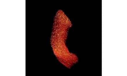
Wang notes that because of its shape and size, it can be challenging to directly detect myelin damage in the spinal cord. The research, he says, “marks the first time we have been able to image its function at the molecular level.”
The release notes that the MeDAS molecular probe operates much like a homing device. It is injected into the body intravenously and is programmed to seek out and bind only to the myelin in the central nervous system. A position-emitting radioisotope label on the molecule allows a PET scanner to detect the targets and quantify their intensity and location. Researchers say the data can then be reconstructed into an image.
Chunying Wu, PhD, first author of the study, instructor of radiology at Case Western Reserve, calls the probe an “indispensible tool” to assist in finding new treatments for MS in the future. “It can also be used as a platform technology to unlock the mysteries of other myelin related diseases such as spinal cord injury,” Wu says.
The university adds that its research team has completed preclinical studies in animals and has begun the process of initiating human trials.
[Source: Case Western Reserve University School of Medicine]