by Talia Konigsberg, PT, DPT
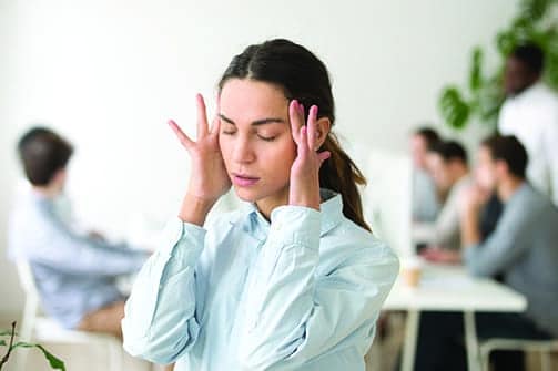
Concussion or mild traumatic brain injury (mTBI) is a pathophysiological process caused by traumatic biomechanical forces that affect the brain. The acceleration deceleration mechanism leads to diffuse axonal injury; it does not necessarily require direct contact to the head. Typically, the recovery from concussion is spontaneous. However, there are those that do not recover within 7-10 days and have persistent symptoms that can last several months or more. Those who have a longer recovery are said to have post concussive syndrome (PCS). According to the ICD-10 definition of PCS, somatic symptoms can include headache, dizziness, general malaise and excessive fatigue, noise intolerance, emotional changes, sleep changes, cognitive impairments including difficulty in performing mental tasks, and memory impairments.1 Because of the diffuse axonal injury and metabolic impairments that occur after mild TBI such as concussion, the impairments are broad.2
The treatment and management of the concussed patient often requires a multidisciplinary approach.3 Symptoms of headaches, sleep disturbances, and emotional changes may require neurological or psychiatric intervention. Cognitive and neuropsychological deficits are a common presentation, and referral to a neuropsychologist for testing and progress monitoring is often necessary. Neuro optometrists or ophthalmologists can test for oculomotor changes and often refer to occupational therapy for vision therapy.
Headfirst Into Therapy
Physical therapy evaluation of a concussed patient is very important in order to create and customize a tailored program to address underlying impairments. The term “dizziness” is commonly reported after concussion but is a vague descriptor that can have various etiologies such as cardiovascular origin, peripheral vestibular dysfunction, post traumatic benign paroxysmal positional vertigo (BPPV), central nervous system disorders involving ocular or vestibular, and cervicogenic origin.2
Post traumatic BPPV is one of the most common forms of peripheral vestibular dysfunction following trauma. Multi canal involvement is more common following trauma, and assessment is crucial.3 BPPV is characterized by episodic vertigo with spinning sensation of oneself or environment. Spontaneous resolution of BPPV can occur, and those with persisting BPPV may report additional complaints of light-headedness or impaired balance. Different positional tests, most commonly the Dix-Hallpike and Roll tests, are utilized to determine the involved canals and direct subsequent maneuvers to treat appropriately and effectively.
Testing and Technology
The direction of the nystagmus during testing will determine which of the semicircular canals are involved. Eighty-five percent of BPPV cases involve the posterior canal, which is indicated by upbeating the torsional nystagmus toward the affected ear lasting less than 60 seconds.2 Testing can be performed in room light or with the use of Frenzel goggles. Frenzel goggles can be beneficial in that fixation is blocked for the patient and the room is dark when placed on the eyes. However, they contain magnifying glasses and a lighting system that is projected on a screen for the clinician to see and monitor more effectively.
Identifying Dysfunction
Visual disturbances are a common symptom report post concussion, and these include blurry or double vision and difficulty focusing. The oculomotor examination can improve the diagnosis of concussion and differentiate between peripheral or central origin and to track progress. Peripheral vestibular dysfunction may include peripheral hypofunction indicated by positive head impulse test, dynamic visual acuity test, or BPPV.2 Central dysfunction may include oculomotor dysfunction, including impaired smooth pursuits, saccades or convergence, motion sensitivity, and/or central vestibular dysfunction that may be indicated by positive vestibular ocular reflex cancellation test or positive spontaneous or gaze-evoked nystagmus. Convergence insufficiency, or the inability to generate enough ocular convergence to maintain clear binocular vision with a near target, can cause dipolpia or blurriness and can be tested with the near point convergence test. The King Devick test can help diagnose slow saccadic movements.5 Headache, fatigue, and loss of concentration are also often reported with oculomotor deficits.2 In the case of central dysfunction, vision therapy and referrals to a neuro optometrist/ophthalmologist and occupation therapy may be indicated.
Posture and Balance
Postural control tests that are commonly used include the Balance Error Scoring System (BESS) and the Sensory Organization Test (SOT). The SOT is a dynamic posturography test of the integration of visual, somatosensory, and vestibular systems responsible for balance and postural stability. Patients with vestibular dysfunction often have increased reliance on visual input for balance.4 The test is comprised of six different sensory conditions: (1) eyes open, fixed support; (2) eyes closed, fixed support; (3) sway-referenced vision, fixed support; (4) eyes open, sway-referenced support; (5) eyes closed, sway-referenced support; and (6) sway-referenced vision and support. A patient’s performance can be compared to normative values and, based on the conditions performed below average, it can be determined which systems are contributing to instability. More recent and advanced technology may incorporate the use of virtual reality for testing and treatment.6
Effects of Exertion
It is not uncommon for a patient with a concussion or post concussive syndrome to report increased symptoms upon exertion. The Buffalo Concussion Treadmill Test (BCTT) is used to diagnose abnormal physiologic response to exercise in concussion and determine exercise tolerance of the patient. The test is not appropriate for all patients, including those with significant balance impairments, cardiovascular disease, pulmonary disease, or orthopedic injuries. The test is administered with a starting speed of 3.6 and an incline of 0. As per protocol, the incline is gradually increased every minute while monitoring rate of perceived exertion, symptom report, and HR. The speed is increased once the maximum incline is reached. The test is stopped with a significant increase in symptoms or when exhaustion is reported. Based on the patient’s threshold heart rate, a customizable aerobic exercise treatment plan can be initiated.1
Origin of Dizziness
Cervicogenic dizziness is not as understood but is commonly a contributor to dizziness following concussion. Cervical involvement may include neck pain, impaired range of motion or passive mobilization of the cervical spine, and impaired neuromotor control of the deep neck flexors.7 The upper cervical spine proprioception plays a role in postural neck reflexes and the cervico-ocular reflex, which complements the vestibulo-ocular reflex. Following acceleration-deceleration injuries to the cervical spine, that cervical proprioceptive input would be impaired and contribute to reports of dizziness and light-headedness. Additionally, these type of injuries can lead to impaired cervical proprioception or kinesthetic sense, which can be tested with the Cervical Joint Position Error Test. The Head-Neck Differentiation test can assist in discerning between vestibular and cervical origin of dizziness.2
Multidisciplinary Matter
When treating patients following concussion, progress with physical therapy will vary with the patient and is often related to the diversity of the symptoms. The physical therapist will often have to prioritize impairments and modify as the plan of care progresses. The multidisciplinary approach is crucial in addressing impairments beyond the scope of physical therapy. RM
Talia Konigsberg, PT, DPT, is a senior PT at Kessler Institute for Rehabilitation in Saddle Brook, NJ. She is a graduate of NYU Doctor of Physical Therapy Program. She has specialized in neuro rehab in both inpatient acute rehab and outpatient. She is a certified vestibular therapist and a member of VEDA (Vestibular Disorders Association). For more information, contact [email protected].
References
1. Leddy JJ, Willer B. Use of graded exercise testing in concussion and return-to-activity management. Curr Sports Med Rep. 2013;12(6):370-376.
2. Herdman S, Clendaniel R. Vestibular Rehabilitation. F. A. Davis Co; 2014.
3. Aligene K, Lin E. Vestibular and balance treatment of the concussed athlete. NeuroRehabilitation. 2013;32(3):543-553.
4. Kim M, Lee DS, Hong TH, Joo Cho H. Risk factor of benign paroxysmal positional vertigo in trauma patients: A retrospective analysis using Korean trauma database. Medicine (Baltimore). 2018;97(49):e13150.
5. Cheever KM, McDevitt J, Tierney R, Wright WG. Concussion recovery phase affects vestibular and oculomotor symptom provocation. Int J Sports Med. 2018;39(2):141-147.
6. Alsalaheen BA Mucha A, Morris LO, et al. Vestibular rehabilitation for dizziness and balance disorders after concussion. J Neurol Phys Ther. 2010:34(2):87-93.
7. Renerker JC, Cheruvu VK, Yang J, James MA, Cook CE. Physical examination of dizziness in athletes after a concussion: A descriptive study. Musculoskelet Sci Pract. 2018;34:8-13.