Computerized eye tracking technology aims to provide objective, thorough assessment for concussion. The technology records eye movement in relation to a simple, circular visual stimulus and produces immediate results showing variability in spatial and temporal pediction.
by Lenore Herget, DPT, MEd, SCS, CSCS
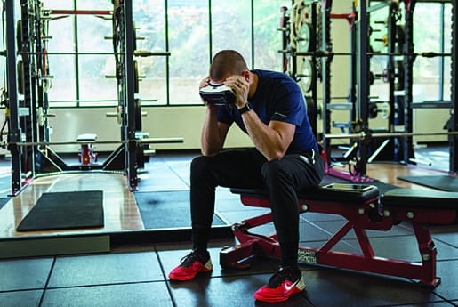
When I examine an athlete after sustaining a concussion, I need to devote ample time to effectively and safely assess many systems in the body that help the athlete perform at top physical and cognitive performance. After collecting a subjective history, including injury mechanism of injury, I do a thorough binocular vision exam. I look at eye teaming skills (convergence and divergence), eye focusing skills (accommodation), and eye tracking skills. Specifically with eye tracking, I assess extraocular muscles with both smooth pursuits (slow, <60 degrees/second) and saccadic (quick, >600 degrees per second) eye movements. Smooth pursuits can be examined with a simple “H”, “I”, and/or “X” test where a target is moved slowly in the formation of the letter and the examiner observes for any nystagmus, difficulty with gaze holding, and accuracy.
A more objective, thorough assessment of this can be done using computerized eye tracking software. This technology, which includes a combination of high-speed cameras and infrared sensors, goes a step beyond a simple smooth pursuit assessment, as it focuses on predictive attention performance and synchronization; both of which can be greatly impaired after concussion. It records eye movement in relation to a simple, circular visual stimulus and produces immediate results showing variability in spatial and temporal prediction. With a .8 test retest reliability, no apparent effort or learning effect and 15 + years of research backing it up, it is a test that has changed the way I practice and manage my concussed athletes as I have tangible, objective data that help identify concussion patterns. And because the test takes only a minute to perform, it can be easily administered on the sideline or in the clinic to help improve diagnostic accuracy and show improvement over time. My athletes have universally taken to this technology and greatly respect the value this technology offers as it shows function in real time, with real numbers and pictures that they can understand and improve upon.
I use this computerized eye tracking software in all of my patients who present after sustaining a concussion; from high school to collegiate to professional athletes and even weekend warriors. The entire process of collecting eye tracking data for each athlete takes about 1 minute, but its quick post-eye tracking analysis and easy-to-interpret numeric and graphed performance scores makes it the most important minute of the examination.
After the functional binocular vision exam, I look at the closely linked vestibular system, for which intact binocular visual function is necessary, and I look at the athlete’s gaze stability or vestibular ocular reflex (VOR), or ability to keep an image centered on the fovea during horizontal and vertical head motion. I do this by measuring how quickly the athlete can move their head side to side and up and down while maintaining focus on a target without symptom provocation. Oftentimes, with concussion, this ability is impaired, and head movement at necessary speeds (2 + hertz for daily activities) causes dizziness, nausea, or blurred vision. Taking a static visual acuity measurement, using a Snellen eye chart or handheld eye chart, will give you a baseline of visual acuity without head motion. Using the same chart, I cue the patient to read the lowest line on the chart that they can while assisting them moving their head side to side, roughly 20 to 30 degrees to each side at 2 Hz. I then repeat the same test with up-and-down head motions. This measurement of dynamic visual acuity should include the difference in line numbers on the eye chart between what they could read without head motion (static visual acuity) and what they could read with head motion (dynamic visual acuity, a measure of VOR). This difference should be no more than one to three lines; less than two in the more elite-level athlete who requires more precise vision with quicker head movements.
A head thrust test is another method to assess for VOR dysfunction; though it is necessary for a practiced examiner to administer. Additionally, the computerized eye tracking software now includes an objective method of measuring VOR, in both the horizontal and vertical planes. The technology includes a metronome or the examiner can use their own device to set the pace at which to measure VOR functioning. Again, this has been very instrumental in objectively assessing how well a patient is doing and in giving them more finite numbers upon which they can improve.
This technology improves my confidence in the diagnosis, and patients value what the data shows. They appreciate being able to have access to real numbers in real time to attribute to their recovery. They likewise are enthused about being able to see improvement charted over time; particularly when baseline scores are available and they can watch their software-generated scores inching closer to baseline.
Prior to using this technology I had a very thorough, effective evaluation for athletes suffering a concussion. I was able to appropriately gather information about the oculomotor, vestibular, musculoskeletal (cervical spine), and cardiovascular systems to generate a functional diagnosis and establish a plan of care to return the athlete safely and efficiently back to play. I had a few examination metrics that I weighted heavier than others as they were more objective and reproducible; and therefore became components of my preseason baseline testing for each team.
When I was first introduced to computerized eye tracking I realized that this would be a game-changer in both the evaluation and the plan of care. After thoroughly reviewing the technology, to the point a “non-techy” like myself can understand, and reading through powerful research studies and decades of data collection, I immediately wanted to be involved.
I was fortunate enough to be an early user of the product and so began incorporating it into my initial evaluations, daily re-evaluations, and return-to-play decision-making. Teams, with which I was already working, were interested in how the quick, reliable, objective data that was being collected and analyzed could be used to more safely care for their athletes.
Additionally, I use this technology to aid in my decision-making to rule out a concussion. When all other oculomotor, vestibular, and neurocognitive testing is normal, and the eye tracking device performance is normal or baseline, I can begin to look elsewhere for symptom generators; most often, the neck. It helps guide earlier treatment in other areas than before using this technology.
When it comes to the rehabilitation side of concussion, we consider other systems not discussed in this paper, such as exercise intolerance/progression and the cervical spine and balance.
From an oculomotor and vestibular standpoint, we use tools that are like the tests and measures listed above to rehabilitate the dysfunctional components. We consider the normative values; for instance, a near point of convergence less than 5 cm, and work on convergence using Brocks strings or two fingers or pencil pushups until the athlete can achieve the norm. There are optometric-approved apps on phones or websites as well that provide screen-based exercises for smooth pursuits, saccades, and tracking dysfunction. Typically, these are worked on at home, progressed in the clinic or training room, and reassessed using the eye tracking technology for improvement.
When VOR is impaired, like the test, we work on improving the reflex until the patient can move their head 20 to 30 degrees to each side at 2 Hz (higher if elite-level athlete) with precise focus (no blurriness) or symptom provocation. That may mean starting at a lower level and making accommodations to achieve perfect visual focus without symptoms; for example, using a larger target in a visually simplistic environment and moving the head at a slower speed (use a metronome to help track progress) until the desired level is achieved. Again, using the eye tracking device periodically to asses for smaller, more objective change can be beneficial for the provider and the athlete as they see positive change that may not be as evident in their day-to-day life or to the naked eye. RM
Lenore Herget, DPT, MEd, SCS, CSCS, is senior physical therapist, board-certified clinical specialist in Sports Physical Therapy, certified strength and conditioning specialist at MGH Sports Physical Therapy, Massachusetts General Hospital, Boston. For more information, contact [email protected].


I had the same question as above. Thanks
What system are you using for the computerized eye tracking?