by Jennifer McWain, MHS, PT; Kelly C. Bonarrigo, PT, DPT; and Kirk Randall, MSPT
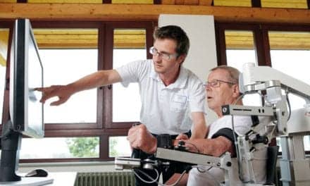
 Kirk Randall, MSPT, Mary Free Bed Rehabilitation Hospital, instructs an outpatient in a strengthening exercise for dorsiflexors.The diagnosis on the prescription that accompanies your next patient says “foot drop.” This can be a misleadingly simple term. Understanding the cause of the foot drop is the key to determining the path of rehabilitation toward recovery, control, or compensation for the foot drop. Table 1 lists common gait abnormalities seen in foot drop. The etiology of foot drop falls into three different categories: neurological, muscular, or anatomic.
UNDERSTANDING THE REHABILITATION MECHANISM
Understanding the recovery mechanism and prognosis is important to providing the best care at the proper time. In upper motor neuron (UMN) lesions, clinicians must take into consideration the acute stage versus chronic stage to direct treatment at restoring control when higher rates of recovery are possible, versus compensating for long-term stable deficits. In the case of lower motor neuron (LMN) deficits, treatment decisions are based on the timing and prognosis for recovery. Disease-related neuropathies may decline while those of a traumatic nature may recover.
Significant dorsiflexion (DF) strength recovery after surgical nerve decompression in cases of foot drop from herniated nucleus pulposus and/or lumbar stenosis was noted in 98% of subjects, with 71% reporting full DF strength recovery.1 In a study of 14 subjects who had foot drop due to damage to the common peroneal nerve of less than 1-year duration and subsequently underwent nerve transfer, 11 subjects had successful restoration of motor grade fair plus to good plus ankle DF; one subject had restoration of fair grade ankle DF; and two subjects had no restoration of DF.2 With nerve transfers, professionals must educate the patient about waiting the approximate 1 mm/day of nerve healing necessary before motor activity can be expected to be seen while compensating for the foot drop in the meantime. Preparing postsurgical patients for reestablishing a safe and efficient gait pattern, no matter what the outcome of surgery, will help them mentally move forward in whatever direction recovery takes them.
Regardless of the cause or prognosis, rehabilitation professionals play a primary role in helping patients return to safe ambulation and transfer status. A primary treatment goal is to improve or maintain ankle DF range of motion (ROM) in both joint and muscle flexibility parameters.3 Typically, patients who have normal or near normal ROM can be taught home stretching programs for the gastrocnemius and soleus muscles. Manual stretching can be used if the patient has neutral or less DF ROM. There are many effective ways to approach stretching from passive to dynamic, and unloaded to loaded positions.4-8 Several studies have shown the use of proprioceptive neuromuscular facilitation produces faster results than typical passive or active stretching alone.6,8,9 Keeping the foot in a subtalar neutral position is helpful for both increasing length and preventing overstretch of the arch.10 Manual therapy for joint mobilization and myofascial stretching also are used to affect contractures at anatomic layers beyond muscle. Application of cold and/or heat has shown benefits for helping patients to relax for more effective stretching.11,12 Serial casting and splints also have their place in providing low load, long duration stretching in more difficult contracture conditions. Charcot joints do not usually improve significantly with splint or casting efforts.13 Medical management of spasticity with delivery of antispasmodic medications via oral or intrathecal routes, and injections of intramuscular neuroparalytic agents such as botulinum toxin or phenol can have significant impacts on the effectiveness of ROM and stretching efforts when plantarflexor spasticity is contributing to contractures.
REBUILDING THE FOUNDATION FOR RECOVERY
Another vital treatment goal is strengthening and neuromuscular reeducation. The role of this treatment is dependent on where the patient is with recovery as opposed to permanent loss of nerve signal. It is best to begin working on goals for rebuilding strength and motor control as soon as possible for a patient who has spinal cord injury and incomplete paralysis, stroke, or head injury, or following surgical decompressions of nerve roots. A patient who has a peripheral nerve injury may be evaluated with surface or needle EMG biofeedback (BFB) for signs of recovery before spending valuable treatment time on this goal.14 Conversely, a patient with progressive multiple sclerosis, adult cerebral palsy without active DF, muscular dystrophy, or peripheral neuropathy may not have strengthening goals.13 There is evidence that strength training can have some slowing effects on the progression of foot drop in Charcot-Marie-Tooth disease.13 There are many methods of strengthening. Surface EMG BFB can help a patient turn the muscle on when they cannot yet produce movement.14-16 BFB has been shown to be helpful with training to turn the muscle on during swing phase through heel strike.17,18 BFB also can be valuable in determining an exercises regime that can help the patient achieve the maximal amount of nerve signal to promote the most efficient strengthening program.14-16 Strength programs will focus on whether a patient can assist in AROM or take resistance. It is particularly important to work strengthening into functional tasks of DF with stepping activities and balance ankle strategies. The success of this goal is proportional to the severity of neural injury and recovery.
Surgical tendon transfer is an option for some patients to regain DF motion control. The most frequent tendon transferred for this purpose is the posterior tibialis tendon.19 Patients who undergo tendon transfer will need specific neuromuscular reeducation to relearn to activate the transferred muscle to its new DF function. Therapists must follow physician protocols for appropriate progression of movement and strengthening regimens. Treatment should retrain the transferred muscle to coordinate the activities of DF during the gait cycle and ankle strategy for balance reactions.
When muscle recovery is delayed or absent, orthotics can provide assistance for positioning the ankle for foot clearance during swing phase and control of plantarflexor moment during stance loading. A custom ankle foot orthotic (AFO) consists of moldable plastic that provides a lightweight product that can be adjusted to ensure even pressure distribution on a patient’s limb. Some patients will prefer a double metal upright brace that attaches to the shoe. Both types can be made with varying degrees of rigidity or flexibility. An articulated AFO or a thinner plastic AFO will allow for a rollover motion during stance phase as it allows for some flexibility into the DF direction. A rigid nonarticulated brace can give knee support to help control buckling and/or hyperextension.
For lower costs—and when the DF movement and knee control needs less assistance—there are multiple options for flexible off-the-shelf products. A posterior leaf spring AFO has minimal plastic and can be heated and molded for a patient. Lightweight carbon fiber AFO manufacturers claim an energy return feature. Elastic straps can be attached from leg to shoe. Stirrup brace products that were developed for sports medicine can be effective at giving the ankle enough support to control mild foot drop.
TREATMENT TREND
A recent trend in the foot drop arena is the use of functional electrical stimulation (FES). FES relies on an intact LMN loop, so it is only effective in the case of upper motor neuron lesions in patients with muscles that respond to electrical stimulation.20-23 Patients need to be screened for contraindications to electrical stimulation including seizure history, internal fixation devices, infections, cardiac pacemakers or defibrillators, cancerous lesions, fractures, and dislocations. Neuromuscular electrical stimulation can be used as a tool to prevent or slow disuse atrophy where muscle strength grades are below a fair grade.20 Patients can be set up to carry out this type of strength program on a daily basis with home use electrical stimulation units.
FES has been incorporated into personal units that are used in place of an AFO. Units on the market today make use of unweighting heel switches or tilt sensors monitoring the angle of the leg to trigger FES placed over the peroneal nerve, anterior tibialis, and ankle evertors to obtain a balanced DF response during swing phase. Patients benefit from developing a use program with these devices under the careful supervision of a trained rehabilitation professional. It is recommended that patients follow specific regimens for building up wearing time, especially when their muscles have been subject to long periods of disuse atrophy. A fatigued muscle can stop responding to the FES, and the patient can suddenly experience a return of the foot drop. Overworking the muscle can cause muscle damage. Patients need to be trained to watch for and respond to skin integrity issues as the result of having electrical stimulation applied for long periods of time.
There have been multiple studies that examine FES orthotics in patients following a stroke.21-28 The results showed a significant improvement in walking speed, cadence, step length, physiologic cost index, ankle ROM, spasticity of calf musculature, Fugl-Meyer scores, and capacity of muscle output when using the FES orthosis along with conventional therapy versus conventional physical therapy alone.22,24 A look at these same types of data based on the subacute versus chronic period after a stroke showed larger improvements were made during the subacute period than the chronic period.23 Therefore, early intervention programs of FES therapy combined with a conventional rehabilitation program may significantly improve the gait and muscle strength in stroke survivors with foot drop. Research subjects with stroke and multiple sclerosis had significant increases in both motor-evoked potential and maximum voluntary muscle contraction after regular use of a foot-drop stimulator.23 This suggested the FES therapy strengthened activation of motor cortical areas and their residual descending connections.
Sometimes the most appropriate response is to surgically fuse the ankle in a functional position with a triple arthrodesis. This is usually a last resort effort as it is most limiting in locking into shoe heel heights and minimizing rollover motion. A Charcot joint will typically end up in a fused position as a progression of the disease. These patients may benefit from rocker bottom shoes. Balance training and assistive devices may be necessary for these patients to safely ambulate.
There is much that can be done to alleviate the problem of foot drop. The approach of the rehabilitation professional is most effective when they consider the problem in the light of defining what might be central, peripheral, stable, progressive, and/or recovering.
CASE STUDY #1
A 56-year-old female suffered a cerebrovascular accident (CVA) with almost complete paralysis of the left side. During her inpatient stay, she was set up with a rigid AFO that had an articulated component that could be cut in, but needed the rigid AFO initially to help with knee control due to buckling and later spastic hyperextension. As hip and knee control improved, the AFO was articulated. About 6 months post-CVA, she trialed and then obtained a FES orthotic, which she used for 3 months. Over this 3-month period, she noticed that when not using the FES, she was able to ambulate on level surfaces at slow paces with some control of the foot drop. She stopped using the FES after 3 months because she began having episodes of “freezing up,” which a neurologist determined to be seizures. When the FES was stopped, she had progressed to just needing DF assist and some mediolateral support, which she was able to obtain with a stirrup brace.
CASE STUDY #2
A 52-year-old female developed foot drop after a right total hip arthroplasty (THA). She also had a 37-year history of left above knee amputation and so compensating for foot drop was extremely difficult for her. She was fit with a posterior leaf spring AFO, trained in a ROM program to prevent loss of DF ROM, and provided monthly surface EMG biofeedback sessions to monitor for progress in neural recovery. When signs of increased motor signal were seen 3 months later, she was trained in a more comprehensive strength training program. By 7 months post-THA, she had functional DF, which allowed her to discontinue use of the AFO. RM
References
1. Girardi FP, Cammisa FP, Huang RC, et al. Improvement of preoperative foot drop after lumbar surgery. J Spinal Disord Tech. 2002;15(6):490-4.
2. Nath RK, Lyons AB, Paizi M. Successful management of foot drop by nerve transfers to the deep peroneal nerve. J Reconstr Microsurg. 2008;24(6):419-27.
3. Katalinic OM, Harvey LA, Herbert RD, et al. Stretch for the treatment and prevention of contractures. Cochrane Database Syst Rev. 2010;(9):CD007455.
4. Page P. Current concepts in muscle stretching for exercise and rehabilitation. Int J Sports Phys Ther. 2012:7(1):109-119.
5. Thacker SB, Gilchrist J, Stroup DF, Kimsey CD Jr. The impact of stretching on sports injury risk: a systematic review of the literature. Med Sci Sports Exerc. 2004;36(3):371–78.
6. Haff GG. Roundtable discussion. Flexibility training. Strength Cond J. 2006;28(2):64–85.
7. Ford P, McChesney J. Duration of maintained hamstring ROM following termination of three stretching protocols. J Sport Rehabil. 2007;16(1):18–27.
8. Sharman MJ, Cresswell AG, Riek S. Proprioceptive neuromuscular facilitation stretching: mechanisms and clinical implications. Sports Med. 2006;36(11):929–39.
9. Swain DP, American College of Sports Medicine. ACSM’s Guidelines for Exercise Testing and Prescription. 7th ed. Philadelphia: Lippincott Williams & Wilkins; 2006.
10. Jung DY, Koh EK, Kwon OY, et al. Effect of medial arch support on displacement of the myotendinous junction of the gastrocnemius during standing wall stretching. J Orthop Sports Phys Ther. 2009;39(12):867-74.
11. Knight CA, Rutledge CR, Cox ME, Acosta M, Hall SJ. Effect of superficial heat, deep heat, and active exercise warm-up on the extensibility of the plantar flexors. Phys Ther. 2001;81(6):1206–14.
12. Brodowicz GR, Welsh R, Wallis J. Comparison of stretching with ice, stretching with heat, or stretching alone on hamstring flexibility. J Athl Train. 1996;31(4):324–327.
13. Sackley C, Disler PB, Turner-Stokes L, et al. Rehabilitation interventions for foot drop in neuromuscular disease. Cochrane Database Syst Rev. 2009;(3):CD003908
14. Basmajian JV. Biofeedback: principles and practice for clinicians. 3rd ed. Baltimore: Williams & Wilkins; 1989:112-113.
15. Dana L, Frank BS, Khorshid L, et al. Biofeedback in medicine: who, when, why and how? Ment Health Fam Med. 2010;7(2):85–91.
16. Brucker BS, Bulaeva NV. Biofeedback effect on electromyography responses in patients with spinal cord injury. Arch Phys Med Rehab. 1996;77:133–7.
17. Bolek JE. A preliminary study of modification of gait in real time using surface electromyography. Applied Psychophysiol Biofeedback. 2003;28:129–38.
18. Woodford H, Price C. EMG biofeedback for the recovery of motor function after stroke. Cochrane Database Syst Reviews. 2006;(2):CD004585.
19. Rath S, Schreuders TAR, Stam HJ, et al. Early active motion versus immobilization after tendon transfer for foot drop deformity: a randomized clinical trial. Clin Orthop Relat Res. 2010;468(9):2477–2484.
20. Pomeroy VM, King L, Pollock A, Baily-Hallam A, Langhorne P. Electrostimulation for promoting recovery of movement or functional ability after stroke. Cochrane Database Syst Rev. 2006;(2):CD003241.
21. Sabut SK, Lenka PK, Kumar R, et al. Effect of functional electrical stimulation on the effort and walking speed, surface electromyography activity, and metabolic responses in stroke subjects. J Electromyogr Kinesiol. 2010;20(6):1170-7.
22. Sabut SK, Sikdar C, Kumar R, et al. Functional electrical stimulation of dorsiflexor muscle: effects on dorsiflexor strength, plantarflexor spasticity, and motor recovery in stroke patients. NeuroRehabilitation. 2011;29(4):393-400.
23. Everaert DG, Thompson AK, Chong SL, et al. Does functional electrical stimulation for foot drop strengthen corticospinal connections? Neurorehabil Neural Repair. 2010;24(2):168-77.
24. Sabut SK, Sikdar C, Kumar R, Mahadevappa M. Improvement of gait & muscle strength with functional electrical stimulation in sub-acute & chronic stroke patients. Conf Proc IEEE Eng Med Biol Soc. 2011:2085-8.
25. Kesar TM, Perumal R, Jancosko A, et al. Novel patterns of functional electrical stimulation have an immediate effect on dorsiflexor muscle function during gait for people poststroke. Phys Ther. 2010;90(1):55–66.
26. Kottink AI, Oostendorp LJ, Buurke JH, et al. The orthotic effect of functional electrical stimulation on the improvement of walking in stroke patients with a dropped foot: a systematic review. Artif Organs. 2004;28:577–586.
27. Sheffler LR, Hennessey MT, Naples GG, et al. Peroneal nerve stimulation versus an ankle foot orthosis for correction of footdrop in stroke: impact on functional ambulation. Neurorehabil Neural Repair. 2006;20:355–360.
28. Stein RB, Chong S, Everaert DG, et al. A multicenter trial of a footdrop stimulator controlled by a tilt sensor. Neurorehabil Neural Repair. 2006;20:371–379.
Table 1. Common gait abnormalities seen in patients with foot drop
Foot scuff or drag during swing phase
Lack of heel strike at initial contact
Foot slap at initial contact
Steppage gait pattern with excess hip/knee flexion during swing phase
Contralateral limb vaulting during ipsilateral swing phase
Ipsilateral hip hike and/or circumduction
Jennifer McWain, MHS, PT, graduated from Grand Valley State University with a BS degree in physical therapy in 1989, and from the University of Indianapolis with a MHS degree with focus on Neurological Physical Therapy in 1993. She has worked at Mary Free Bed Rehabilitation Hospital in Grand Rapids, Mich, for 21 years in inpatient therapy, outpatient therapy, pediatric and adult spasticity management clinics, and the adult amputee clinic. She is also the primary physical therapist with the neuromuscular reeducation biofeedback program and amputee programs.
Kelly C. Bonarrigo, PT, DPT, graduated from Duquesne University in 2005 with a bachelor’s degree in health science and a bachelor’s degree in biology. She also earned a doctorate in physical therapy from Duquesne University in 2007. Bonarrigo is currently employed at Mary Free Bed Rehabilitation Hospital on the inpatient stroke team.
Kirk Randall, MSPT, graduated from Grand Valley State University in 1993. Randall is currently employed at Mary Free Bed Rehabilitation Hospital. He is the primary PT with the outpatient stroke program. Randall has expertise in both inpatient and outpatient orthopedics and neurological rehabilitation. For more information, contact [email protected].